Extracorporealshock wave lithotripsy machines
A lithotripsy machines have basically 4 basic components:
(1) A Shockwave Generator
(2) A Focusing System
(3) A Coupling Mechanism
(4) An Imaging/Localization Unit.
Shockwave Generator
Shockwaves are generated using electro hydraulic principle, this is the original method of shockwave generation, electro hydraulic, means that the shockwave is produced via spark-gap technology. In an electro hydraulic generator, a high-voltage electrical current passes across a spark-gap electrode located within a water-filled reflector. The discharge of energy produces a vaporization bubble, which expands and immediately collapses, thus generating a high-energy pressure wave.
Focusing Systems (Reflector)
The focusing system is used to direct the generator-produced shockwaves at a focal volume in a synchronous fashion. The basic geometric principle used in most lithotripters is that of an ellipse. But in lithotripters manufactured by TMC Europa Ltd., a further advanced geometry of dual focus ellipso-paraboloid is used. Shockwaves are created at one focal point (F1) and converge at the second focal point (F2). The target zone, or blast path, is the 3-dimensional area at F2, where the shockwaves are concentrated and fragmentation occurs.
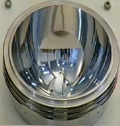
Focusing Reflector
Coupling Mechanisms
In the propagation and transmission of a wave, energy is lost at interfaces with differing densities. As such, a coupling system is needed to minimize the dissipation of energy of a shockwave as it traverses the skin surface. The usual medium used is water, as this has a density similar to that of soft tissue and is readily available. In TMC ESWL systems a small water-filled cushion with a silicone membrane fitted in front is used to provide coupling between the shockwave head and the patient. A small amount of Ultrasound Gel or Silicone Oil is used to provide air-free contact.

Dry Coupling for Lithotripsy
Localization systems
Imaging systems are used to localize the stone and to direct the shockwaves onto the calculus, as well as to track the progress of treatment and to make alterations as the stone fragments. Fluoroscopy, which is familiar to most urologists, involves ionizing radiation to visualize calculi.
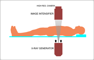
Localization with Fluoroscopy
As such, fluoroscopy is excellent for detecting and tracking calcified and otherwise radio-opaque stones, both in the kidney and the ureter. Conversely, it is usually poor for localizing radio lucent stones (e.g., uric acid stones). To compensate for this shortcoming, intravenous contrast can be introduced or (more commonly) cannulation of the ureter with a catheter and retrograde instillation of contrast (i.e. retrograde pyelography) can be performed.
Extracorporeal shock wave lithotripsy for Gallstones
Gallstones form in the gallbladder, they can be as small as a grain of sand or as big as a golf ball. Gallstones are hard, pebble-like deposits, most commonly made of cholesterol or else made of bilirubin.

Gallbladder stones do not have alternative treatment options other than surgery. ESWL or lithotripsy procedure is rarely performed on gallstones, it is sometimes used on patients having chronic cholecystitis (inflammation of gall bladder) or who are not strong enough or fit for surgical procedure.
ESWL from TMC
Model TMC-IA

The TMC-IA ESWL System: ALSO CALLED AS THE SMALL GIANT, THE TMC-IA IS MODULAR AND ALL IN ONE COMPACT UNIT, DESIGNED PRIMARILY FOR SMALL CLINICS AND PORTABLE USE. WITHOUT AN IMAGING SYSTEM.
Model TMC-SA

The TMC-SA ESWL System: THE TMC-SA IS A SUPERIOR AND VERSATILE LITHOTRIPSY SYSTEM DESIGNED TO BE USED WITH MOST OF THE FLUOROSCOPY C-ARM UNITS AVAILABLE WITH THE CLINIC OR HOSPITAL.
Become a dealer
If you are interested in becoming a TMC dealer in your country or state, Just click the button below, and we will initiate a dealer appointment process...
Model compact

The TMC-Compact, as its name describes, is a small compact unit which has a very small footprint as compared to other ESWL therapy systems. All the subsystems are integrated into one unit and no special arrangements are required to perform ESWL therapy.
Model TMC-X4

TMC-X4 is a ground-up revolutionary, multi functional with ease of operation ESWL equipment. With hugely flexible, customizable and extendable far into the future. A device you would love to own.
Use the form below to send us a message.
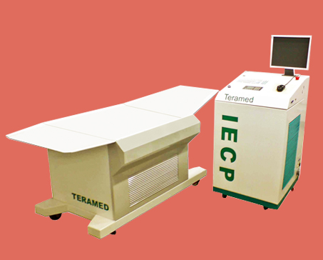
TMC's IECP Devices Increased external counterpulsation equipment
For More detailsClick on the device of your interest

IECP Model TMC - Brisk